Cancer Diagnosis, Accelerated by AI
January 31, 2026 | Saturday | Analysis | By Ayesha Siddiqui
Cancer remains a leading cause of death worldwide, with Asia bearing a disproportionate burden. In 2020, the region accounted for nearly half of global cancer cases and over 58 per cent of cancer-related deaths. Lung, breast, and colorectal cancers were the most commonly diagnosed. Artificial intelligence (AI) has shown particular promise in early detection, where timely diagnosis can significantly improve outcomes. Today, AI supports cancer diagnosis across medical imaging, digital pathology, and genomic analysis, helping clinicians review scans, analyse tissue samples, and interpret complex data more efficiently. Studies show that AI can detect suspicious findings earlier than routine clinical review in some cases. In China, systems such as Alibaba’s DAMO GRAPE and Huawei’s RuiPath have demonstrated high diagnostic accuracy and are already deployed in clinical settings. Beyond detection, machine-learning models are increasingly used to predict treatment effectiveness and patient outcomes. As World Cancer Day is observed on February 4, attention is turning to whether the rapid deployment of AI tools across the Asia-Pacific region reflects genuine clinical impact or continued technological hype.
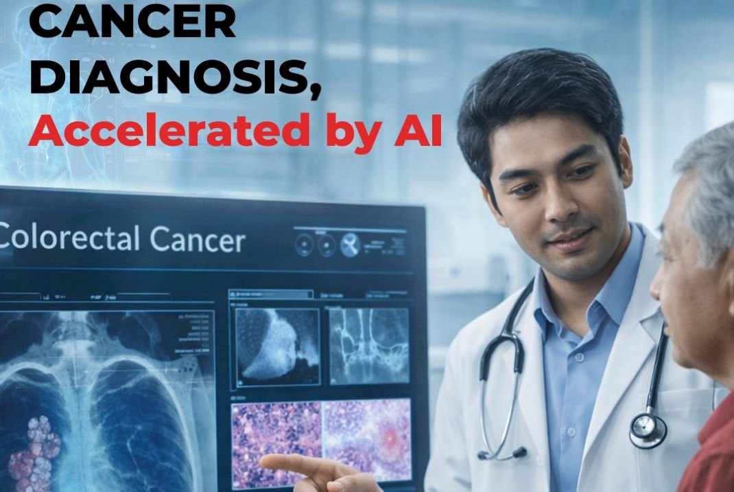
image credit- shutterstock
Cancer remains one of the leading causes of death globally, with Asia carrying a disproportionate share of the burden. In 2020, the region accounted for 49.3 per cent of global cancer incidence and 58.3 per cent of global cancer mortality. The most commonly diagnosed cancers were lung cancer (13.8 per cent), breast cancer (10.8 per cent), and colorectal cancer (10.6 per cent) according to the GLOBOCAN 2020 report.
Early detection is where artificial intelligence (AI) has shown the greatest potential. AI is now used across several stages of cancer diagnosis, including medical imaging, digital pathology, and genomic and molecular analysis. It helps clinicians review scans, analyse tissue samples, and interpret large datasets more efficiently, and in some settings supports risk assessment and clinical decision-making.
Various studies show that AI can identify suspicious findings at an early stage, in some cases before they are detected during routine clinical review. In China, Alibaba’s DAMO GRAPE system for gastric cancer screening outperformed human radiologists, achieving 85.1 per cent sensitivity and 96.8 per cent specificity, and has been deployed in provinces including Zhejiang and Anhui. Huawei’s RuiPath platform supports pathology workflows at Shanghai’s Ruijin Hospital, reducing diagnosis time to seconds, while AI-enabled lung cancer multidisciplinary systems have reached around 90 per cent diagnostic accuracy with high clinician adoption. By mid-2025, these systems had screened more than 180,000 imaging scans. Beyond detection, findings from the RADIOHEAD prospective longitudinal study published in 2025 showed that machine-learning models can better predict treatment effectiveness and patient outcomes in advanced non-small cell lung cancer, with potential applicability to other cancer types.
It is worth noting that the US Food and Drug Administration (FDA) has approved more than 70 AI-associated devices, with 54.9 per cent focused on cancer radiology and 19.7 per cent on pathology. By indication, the largest share of approved or clinically used AI tools target general cancer detection (33.8 per cent) and breast cancer (31.0 per cent), followed by lung cancer (8.5 per cent) and prostate cancer (8.5 per cent) according to a Cancer Journal report.
As World Cancer Day is observed on February 4, we look at who is developing and deploying AI tools for cancer diagnosis across the APAC region, how widely they are being used today, and whether their use reflects real clinical impact or ongoing hype.
Asia-Pacific AI Cancer Diagnostics Landscape
In the Asia-Pacific region, there are more than 110 oncology informatics startups, according to data from Tracxn. South Korean companies are leaders in this space. Chief among them is Lunit, which develops AI software for cancer screening and diagnosis across radiology and pathology. Lunit works with hospitals, screening programmes, and healthcare providers across Asia, Europe, the Middle East, and North America to deploy its AI solutions in routine clinical settings, including population-level screening and hospital-based diagnostic workflows.
Another major player is Coreline Soft, which has played a central role in population-scale lung cancer screening in South Korea. Since 2017, the company has operated the National Lung Cancer Screening Quality Management and Information System, providing nationwide quality assurance across screening institutions for nine consecutive years.
Other companies including Vuno develop AI solutions for medical imaging and clinical data analysis across multiple disease areas, Deep Bio focuses on AI-driven digital pathology to support cancer diagnosis and prognostication. At the national level, the National Cancer Center AI team continues to work on integrating AI with clinical data to support cancer research and deliver diagnostic assistance services, further strengthening South Korea’s position at the forefront of AI-enabled oncology.
Australia is also active in AI-enabled cancer diagnostics, with a mix of commercial companies and large-scale research initiatives. Among commercial players, Annalise.ai develops AI software to detect a wide range of findings in chest X-rays, supporting high-volume diagnostic workflows. IntervalRisk provides AI-based breast cancer risk assessment using mammography images, while AlleSense commercialises NanoMslide, a nanotechnology- and AI-enabled biosensor for cancer cell detection. Major research and implementation initiatives include BRAIx, a multi-institutional Victorian programme involving the University of Melbourne, St Vincent’s Institute, BreastScreen Victoria, and the Australian Institute of Machine Learning to enhance breast cancer screening. Australia has also integrated Lunit’s AI into BreastScreen NSW, and researchers from the University of Melbourne and the Skin Health Institute are developing a handheld AI- and thermal imaging–based device for real-time skin cancer detection.
China has a growing number of big technology companies and medical AI firms working on AI-enabled cancer diagnosis. Alibaba DAMO Academy has developed PANDA (Pancreatic Cancer Detection with Artificial Intelligence), an AI system for identifying early-stage pancreatic cancer from non-contrast CT scans, which has also been extended to liver and gastric cancer detection. Tencent has developed medical AI tools for the early diagnosis of oesophageal, lung, and breast cancers using clinical datasets from partner hospitals. In addition to large technology firms, companies such as Deepwise Technology have received regulatory approval for AI-based mammography screening software. Other companies active in this area include Pharus Diagnostics, VoxelCloud, LinkDoc, and Baishi Medical Technology.
Singapore is also active in the region, with companies deploying AI tools across pathology and medical imaging. Qritive, in collaboration with Roche, has deployed AI-driven prostate cancer grading on Roche’s digital pathology platform, supporting visual segmentation and improved workflow efficiency for pathologists. FathomX has developed FxMammo, a radiological computer-assisted detection and diagnostic software approved by Singapore’s Health Sciences Authority. FxMammo supports breast cancer detection in mammography by identifying suspicious image-based features and is intended for use as an adjunct tool to assist physicians in clinical decision-making.
At the national and research level, Project RAPIER (Radiology Pathology Information Exchange Resource) aims to create a comprehensive radiology-pathology data lake focused on liver lesions. The initiative supports the development of deployable AI-driven applications capable of detecting liver abnormalities, describing imaging features, and assisting in diagnosis. These efforts are expected to catalyse next-generation AI-enabled clinical decision support, predictive analytics, and precision medicine. The recently launched Research Institute for Cancer Prevention Screening and Early Detection (RISE) focuses on developing less invasive and more accurate cancer screening and early detection tools, reinforcing Singapore’s commitment to innovation in preventive oncology.
India is also seeing growing activity in AI-enabled cancer diagnostics, with startups focusing on early detection, imaging, and prognostic testing suited to high-volume clinical settings. NIRAMAI Health Analytix uses AI-driven thermal imaging to enable non-invasive breast cancer screening without radiation, supporting earlier detection in both urban and rural settings. 1Cell.Ai applies AI to single-cell analysis to detect circulating tumour cells from blood samples, aiding early diagnosis and disease monitoring. Qure.ai develops deep-learning tools for medical imaging, with multiple FDA-cleared solutions used globally for lung cancer detection and workflow triage. In addition, OncoStem Diagnostics has developed CanAssist Breast, an AI-powered, proteomics-based test that predicts five-year breast cancer recurrence risk to guide treatment decisions, while companies such as SigTuple and Accubits Invent are applying AI to pathology and diagnostic decision support.
Japan is also strengthening its push into AI-enabled cancer diagnostics, with companies focusing on early detection in clinical settings. AI Medical Service was founded to address gaps in gastrointestinal cancer detection by applying AI to endoscopic imaging, supporting earlier identification of lesions during routine procedures and aiming to improve outcomes through earlier diagnosis. Craif, based in Nagoya, is focused on making early detection more routine as a way to improve cancer survival rates. The company has developed miSignal Scan, a urine-based cancer screening test that uses its Bio-AI platform to analyse microRNA patterns associated with cancer, enabling detection before symptoms appear. Craif is also developing a Bio-AI-enabled medical device for the early detection and accurate diagnosis of pancreatic cancer, a disease with limited screening options, with development programmes underway in both Japan and the United States.
Apart from private companies, several research institutions across the region are also developing AI tools, often working directly with hospitals to test algorithms in real clinical settings and support prospective validation studies.
How Widely Is AI Actually Being Used Today
Perhaps the most sought after area for AI after drug discovery has been cancer diagnostics. 80 per cent of AI-associated devices approved by the FDA for oncology are focused on diagnostics. In Asia Pacific, market estimates show that the AI cancer diagnostics market generated about $41.7 million in 2023 and is projected to reach around $247.4 million by 2030, accounting for roughly 21.5 per cent of the global AI cancer diagnostics market in 2023, according to the Grand View research report.
“Machine learning – a key subset of AI – has been used by many companies in the diagnostics space for over 10 years. Companies use AI to drive insights into target discovery, treatment effectiveness prediction, and diagnosis,” said Simranjit Singh, Chief Executive Officer, Guardant Health AMEA. Guardant Health uses machine learning with methylation profile data to identify cancer-specific genomic patterns for detection and prediction, supported by published and validated algorithms. The company partners with ConcertAI to combine real-world clinical data with genomic and epigenomic insights, supporting biopharma clinical study design and cancer therapy development. It also works with ZephyrAI to apply AI in identifying novel cancer biomarkers for drug development, targeted therapy selection, and treatment response monitoring.
AI has evolved into a central component of the modern diagnostic ecosystem, particularly through its integration into digital pathology and imaging workflows. Today, AI is commonly used to automate routine tasks such as cell counting and tissue quantification, allowing pathologists to focus on more complex cases and decision-making.
“AI is already present in cancer diagnostics, but its routine clinical use remains selective and uneven. Digital pathology and radiology have seen early success, particularly for tasks such as image pre-screening, region-of-interest detection, cell counting, and quality control,” said Sebastian Grote, CCO, X-ZELL GROUP. X-ZELL is pioneering the field of next-generation cytology. The company’s mission is to transform cancer diagnostics by fusing novel laboratory technology with digital imaging to detect and visualise individual atypical cells in minimally invasive body liquids and digitise them for instant on-screen analysis.
As these applications mature, AI is increasingly being positioned less as an experimental tool and more as part of the core diagnostic infrastructure. “AI now sits inside cancer diagnostics as a cost, capacity, and standardisation tool, which is why its use has become difficult for health systems to avoid. In liquid biopsy and genomic workflows specifically, AI-supported bioinformatics turns high-volume sequencing data into clinically actionable results fast enough to influence treatment decisions, and consistently enough to support system-wide standardisation,” said James Lumsdaine, CEO, Avitia. Canada-based startup Avitia, an artificial intelligence (AI)-powered cancer diagnostics company advancing access to fast and affordable molecular testing, has announced its entry into Thailand through a partnership with The Chitrapatima Foundation.
Adoption patterns also vary by geography. “AI is increasingly integrated into cancer diagnostics in China, particularly for early detection of gastric, pancreatic, and lung cancers through routine CT scans and pathology analysis. In China, AI is mostly used to aid radiologists in detecting subtle early-stage lesions on routine imaging, such as lung nodules, microcalcifications in mammography, and small gastric/pancreatic tumors on non-contrast CT scans,” said Ming Yii Lai, Senior Consultant, Daxue Consulting.
Other APAC countries are also following suit. “We are seeing increased interest across the Asia-Pacific region, where digital pathology algorithms can help standardise diagnostic quality in high-volume laboratory environments. These computation digital pathology algorithms are computer models developed using deep learning and other advanced machine learning techniques to recognise specific features within digitised, whole-slide images. Computational digital pathology algorithms can aid pathologists by pre-screening slides to flag potentially relevant cases for review, streamlining the prioritisation of workloads in high-volume laboratories or performing quantification and calculations that the pathologist cannot, to generate diagnostic insights. For example, one image analysis algorithm for non-small cell lung cancer is currently in use across hospitals and laboratories in India, South Korea, Malaysia, and Vietnam, supporting PD-L1 scoring to inform immunotherapy decisions. In one large Malaysian laboratory network, full digital pathology adoption alongside AI reduced turnaround times from nearly a week to three days, while increasing slide processing capacity ten-fold,” said Christopher Chiam – Head of Digital Health Solutions, Roche Diagnostics Asia Pacific.
Beyond pathology, similar approaches are being applied to liquid biopsy and molecular diagnostics, with national health system integration emerging as a key focus. “Our company recently entered Thailand, which has a similar national health system set-up as Canada, with the objective of improving cancer care through cost-effective liquid biopsy testing and AI/ML-powered bioinformatics. This is being done in partnership with The Chitrapatima Foundation, which develops projects to improve the well-being of the Thai people,” said Lumsdaine.
They are establishing Thailand’s first advanced liquid biopsy cancer testing ecosystem for hospitals and communities, leveraging AI-powered precision diagnostics to provide Thai people with precise treatment guidance through a simple blood test, regardless of their location within the country.
“Medical AI is rapidly evolving from a ‘point accuracy’ competition to a vital ‘productivity infrastructure’ for national screening systems. Its true value lies in systemic productivity: not only finding cancers earlier but also reducing the radiologist’s workload by up to 70 per cent , standardising long-term follow-up protocols, and extracting multi-disease risk profiles from a single LDCT scan. This operational shift is what ultimately makes the large-scale expansion of national screening programmes both feasible and sustainable,” said Youna Kim, Deputy General Manager (PR Team), Coreline Soft, South Korea.
This also has direct implications for access to care. “AI is a key part in broadening access to cancer care. It increases workflow efficiency and precision by saving the oncologist from having to analyse raw results and integrate massive amounts of information. By allowing centers that do not have in-house bioinformatics expertise to offer testing on premise, AI-driven solutions are also helping to increase access to cancer testing by moving it out of major hospitals and cancer centers to a wider range of hospitals closer to patients. This reduces the geographic and economic barriers to access,” said Lumsdaine.
Is It Mostly Hype?
Although numerous studies highlight improvements in accuracy, efficiency, and workflow enabled by artificial intelligence, experts have mixed opinions on its real-world impact in cancer diagnosis.
Some experts think it is incremental. “AI in cancer diagnostics is now embedded in daily workflows, especially in medical imaging. However, real-world impact has been incremental, partly because models must constantly adapt to variations in scanners and acquisition protocols. These domain shifts dilute performance gains outside tightly controlled trials,” said Prof. Terence Wong, Associate Professor, Department of Chemical and Biological Engineering at HKUST. At HKUST, his team’s CHAMP Microscope tackles this by integrating the imaging hardware and AI software into a single, co-designed device, enabling robust, label-free histology in minutes for on-the-spot cancer visualisation with far less variability—an approach that could be truly transformative and broadly deployable across settings.
Others echo this assessment, arguing that progress to date has been real but bounded by structural constraints. “So far, AI’s impact has been largely incremental rather than transformative. The evidence shows improvements in efficiency, reproducibility, and sensitivity for well-defined tasks, such as detecting rare cells or reducing inter-observer variability. For real clinical cut-through, continued progress in digitisation and standardisation at the front end of the diagnostic workflow is essential to generate rich, reproducible data that is ready for AI. This is where technologies such as multiplex staining and high-speed imaging become critical. If we get these fundamentals right, AI will initially deliver value not as a replacement for expert cytopathologists, but as a force multiplier – unlocking the full potential of visual biomarker and morphology data to enable more confident, standardised, and clinically actionable diagnoses. That type of data is inherently well suited to machine learning, which is why I see substantial long-term potential for AI when implemented in the right way,” said Grote.
Lumsdaine agrees, “Despite the advances, the real-world impact of AI on diagnostic outcomes has remained largely incremental. The primary constraints have not been technical, but structural: slow clinical rollout due to fragmented integration into care pathways and unresolved reimbursement in many cases.”
However, evidence from scaled implementations suggests that when these barriers are addressed, AI can deliver measurable clinical and system-level value. “Our company recently published a real-world study in Current Oncology (MDPI) detailing our pan-Canadian ACTT liquid biopsy initiative, which used the company’s AI/ML bioinformatics platform to bring our Follow It LBx panel across Canada. The published results clearly indicated a significant benefit to patients for timely selection of appropriate therapy and demonstrated strong potential for system-wide cost-savings. ACTT was launched during COVID-19 to relieve diagnostic bottlenecks and then expanded to address longer-term systemic delays in cancer diagnostics, delivering high-quality, actionable results for 97 per cent of participants within an average 8-day turnaround. The initiative served more than 4,000 patients nationwide, including people in rural and remote regions. Independent studies from three major Canadian institutions demonstrated that the use of Avitia AI/ML-driven LBx solution enabled ~50 per cent reduction in time within the diagnostic pathway and provided additional actionable information relative to standard-of-care testing (47 per cent vs. 18 per cent ). These benefits underpin critical improvements in cancer care by allowing patients to receive the most appropriate therapy faster and more often than in the absence of such tools,” said Lumsdaine.
However, some large diagnostics players view AI’s impact as already crossing the threshold from incremental improvement to transformation, particularly in areas where consistency and precision directly influence treatment access.
“The impact of AI at Roche is viewed as transformative because it addresses the critical challenge of diagnostic consistency and precision. Early evidence shows that AI-enabled diagnostics can identify subtle biomarkers and morphological patterns that may be imperceptible to the human eye, potentially leading to significantly earlier and more accurate detection. By reducing inter-observer variability, AI ensures that a patient’s diagnosis is not dependent on which specialist reviews their case, a meaningful leap forward for health equity. For example, using AI algorithms to increase the accuracy of detecting PD-L1 expression in lung cancer ensures that a patient’s diagnosis and treatment eligibility are less dependent on where they are treated or which specialist reviews their case. Ultimately, AI is shifting the needle from reactive treatment to proactive precision medicine, and may directly influence long-term patient survival,” said Chiam.
This view is reinforced by advances in early detection and disease monitoring. “AI has enabled us to enhance the sensitivity of tests to detect early-stage cancers, allowing for early intervention that is well known to improve outcomes. It also allows us to detect minimal residual disease and monitor for cancer recurrence in patients with early-stage cancer. In advanced cancer, AI also enabled us to expand biomarker identification and introduce molecular subtyping capabilities to help oncologists identify optimal treatment plans,” said Singh.
Whether AI ultimately reshapes cancer diagnosis at scale will depend on how effectively health systems and regulators integrate these tools into routine care. “In other words, the question is no longer whether AI gets used — it is whether health systems can afford to deliver timely, equitable precision diagnostics without it,” Lumsdaine sums it up.
Ayesha Siddiqui